Chitosan Nanoparticles - Sophie Blee-Goldman, Sakshi Shah, & Faith Lemire-Baeten
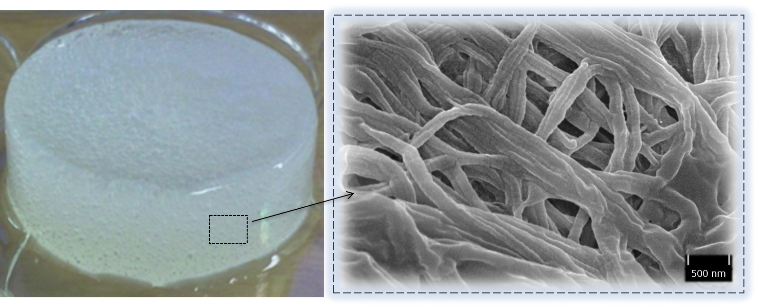
Figure 1: Two views of the patch engineered by Orwin Lab's Brain Patch team. The collagen gel scaffold as seen by the naked eye is shown on the left, while the fibrous collagen matrix as seen on the SEM is shown on the right. The Brain Patch has three primary components: collagen gel (which provides the mechanical support), mesenchymal stem cells (which differentiate into brain cells and help with tissue loss during TBI), and the chitosan nanoparticles (which provide antibacterial and anti-inflammatory properties that fight external infection as well as damage due to the brain's secondary inflammatory response).
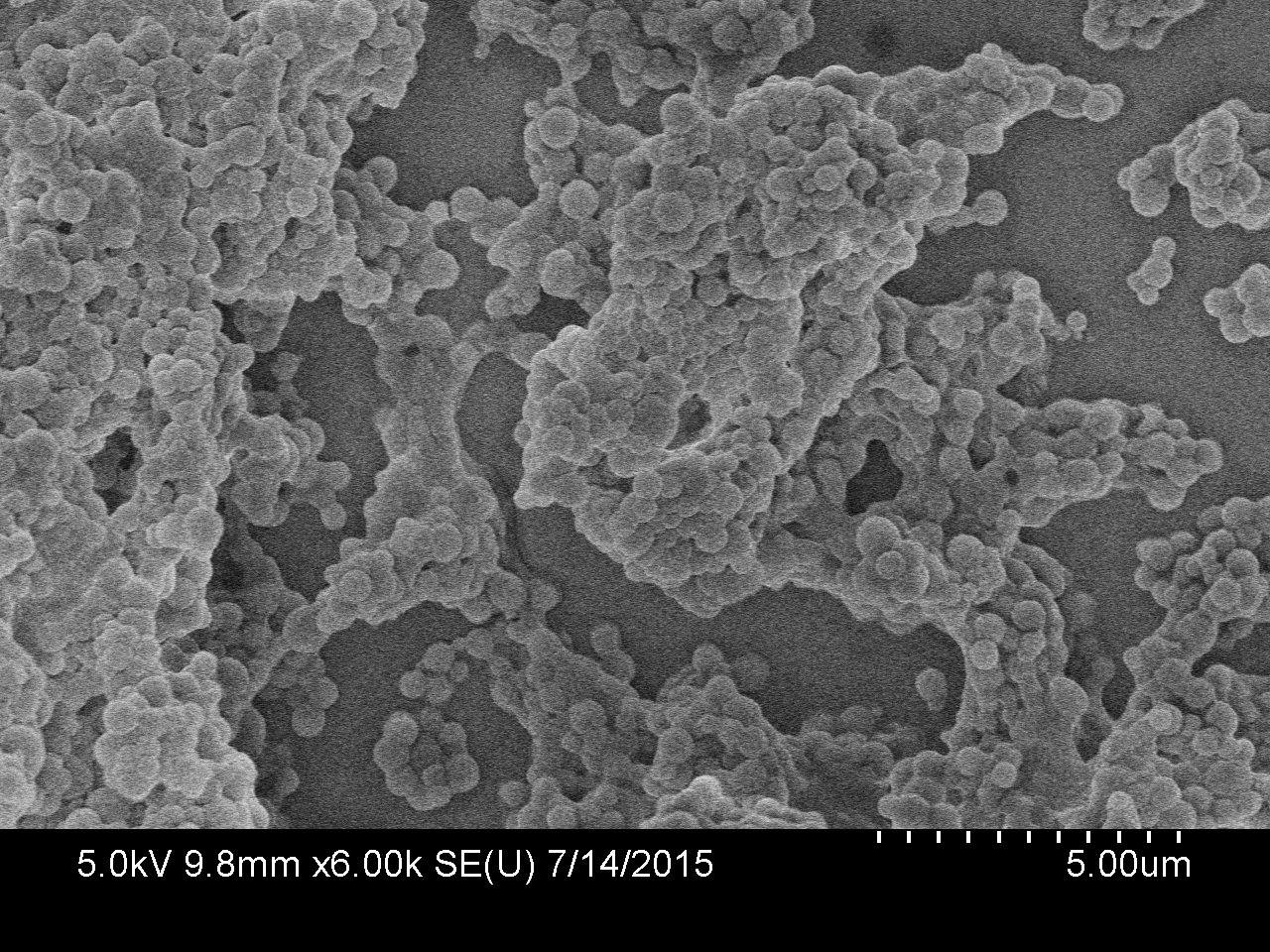
Figure 2: An image taken on the SEM at Pomona College of the chitosan nanoparticles produced in our lab. This sample was collected by impaction on a silicon slide, and using software to analyze their size the average particle diameter was found to be and were analyzed by software to have an average diameter of 441.9 ± 123.6 nm.
Overview
This project is in collaboration with Prof. Liz Orwin's group in the engineering department at Harvey Mudd. The goal of our work is to produce chitosan nanoparticles, whose antibacterial properties are essential for Orwin Lab's "brain patch" (Figure 1), a tissue-engineered treatment for Traumatic Brain Injury (TBI).
Current Progress
We synthesize nanoparticles through a two-stage process of atomization and rapid evaporation. An ultrasonic atomizer, or nebulizer, allows us to produce an aerosol of micron-scale droplets from a solution containing dissolved, commercially-available chitosan. These droplets are then directed through a heated environment where they rapidly lose water. As the solvent evaporates the strands of chitosan polymer contained in each droplet are compressed together and ultimately form spherical, dry nanoparticles of chitosan.
The collection of these airborne particles exiting the evaporation stage is a current area of focus. Previous methods tested include dry inertial impaction (used to obtain the sample in Figure 2) as well as "wet impaction" with a Particle Into Liquid Sampler (or PILS, borrowed from Prof. Leila Hawkins in Harvey Mudd Chemistry). Recently, we have been successfully using a Pall Laboratory nylon membrane filter. Initial results suggest this approach results in high collection efficiency with limited contamination. It also collects the particles in such a way that they are easily recovered from the filtration device in a quantifiable amount. These are all problems we have encountered with previous collection attempts, so we hope to begin producing enough for antibacterial testing and general integration with the Brain Patch Team soon!
(NOTE: Clicking any of these links will go to an embedded PDF screen. To get back to this page, simply click the back button on your browser.)
If you would like to see the slides from a presentation that Sophie gave recently for her senior fall 2015 thesis talk,
click here.