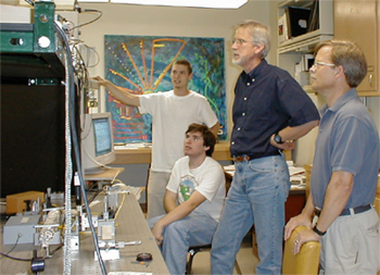
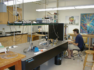
 Harvey Mudd College's optical coherence microscope (OCM) project is a highly interdisciplinary
approach to research in developmental biology. Funded primarily by a 1996 NSF award, the OCM
project has involved undergraduate students from all major academic programs at the college, as well as
5 faculty from 3 academic departments.
Over 50 students and faculty from HMC alone have participated in collaboration with others
at the California Institute of Technology, University of Colorado State,
and Beckman Laser Institute Medical Center, since the project's beginning in 1995.
The OCM research experience has
promoted scientific curiosity and enthusiasm in many students, while also
expanding the research tools available to faculty beyond their traditional disciplinary
boundaries.
Optical coherence microscopy has potential for application in developmental biology in its ability to image tissue regions that are inaccessible to conventional microscopes. The high light scattering properties of biological tissue renders such techniques as two-photon and confocal microscopy useless for imaging much beneath the surface layer of tissue. Optical coherence microscopy overcomes this limitation, allowing three-dimensional imaging of cells or groups of cells at depths of up to 1 millimeter below the surface. The design of the instrument allows imaging of biological samples non-destructively and non-invasively, allowing processes to be monitored in vivo. Taking advantage of modern visualization software, our approach to optical coherence microscopy provides a method for constructing three-dimensional time-lapse movies of tissue development, a clear advantage for developmental biologists.
 Currently two model organisms are studied with the microscope:
the frog
Xenopus laevis and the plant Arabidopsis thaliana. In collaboration
with researchers and faculty in the Biological Imaging Center of Dr. Scott Fraser at
the California Institute of Technology, extensive studies of frog gastrulation
have been performed using the OCM. Andrew Schile (HMC 2001) focused his studies on this process,
constructing numerous time-lapse movies of
gastrulation in the frog. Dr Mary Williams of the HMC Biology department
has coordinated plant imaging in collaboration with Dr. June I. Medford and others at Colorado State University.
These studies are focused on the dynamic process of leaf formation (phyllotaxis) during
plant development. In particular, we are studying the
initiation of leaf primordia at the shoot apical meristem.
The success of the OCM project has led to a number of published articles on the development of the OCM instrument and the results of biological studies. These articles, as well as some theses papers from seniors involved in the project, are available in full text on the publications page. In addition to use in developmental biology, our 3-dimensional approach to optical coherence microscopy has possible clinical applications in dentistry, dermatology, opthalmology, and endoscopic medicine. It is also possible that the OCM will be implemented in the agricultural industry to develop more productive and nutritious crops.